Characteristic Beam Patterns of X-ray Include Which of the Following
The actual size of the largest focal spot is no more than a few millimetres in diameter. Mm 3 where P v h and t are the beam power W beam velocity mms hatch spacing or line offset mm which is the spacing between melt tracks and layer thickness mm respectively.

54 Questions With Answers In X Ray Imaging Scientific Method
What are 7 properties of xrays.
. Longer x-rays are fewer in frequency less waves throughout the beam and produce a beam that is less penetrating that shorter x-ray waves. The Electron Column 13 31 Beam Current 14 32 Accelerating Voltage 14. X-rays travel in straight lines and a beam of X-rays diverges from its source.
The Primary Xray Beam Xrays are formed within a very small area on the target anode called a focal spot. Structures the beam hits first will be magnified in relation to those which are nearer the detector. They affect photographic emulsions.
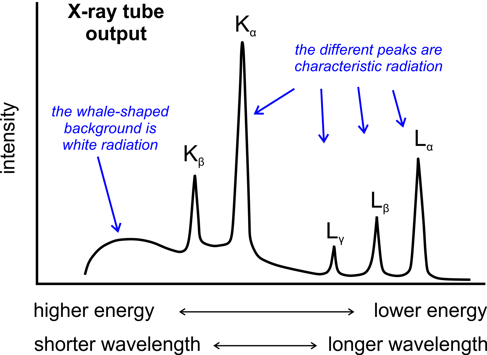
Typically round collimators are built into open-ended aiming cylinders. Cross section of xray beam is called the radiation field. Continuous and Characteristic X-ray Spectra When the target material of the X-ray tube is bombarded with electrons accelerated from the cathode filament two types of X-ray spectra are produced.
X-Ray beams that are parallel with wide projection of the filament have a focal shape of a line. For example the quality of a γ-ray beam emitted from a 60 Co source can be stated in terms of 117 and 133 MeV average 125 MeV or simply cobalt-60 beam. Namely they have velocity wavelength frequency and amplitude.
Figure 136 presents the beam patterns of the standard LCMV scaled LCMV modified LCMV and the SU rate maximization approach. A lead apron should be carried with the unit and worn by the radiographer during exposure. 2 The K-shell electron is removed only if the energy of the incident electron is greater than the K-shell binding energy leaving a vacancy in the K-shell.
1 E a P v. The tube voltage kVp tube current mA and filtration all influence the quantity and quality of the x-ray beam but not the specific energy of characteristic photons. Many materials are transparent to x-rays.
The distance between the beam exit and entrance on the x-ray film. Shape of the x-ray beam and therefore the volume of irradiated tissue within the patient. Free air in the abdomen taken with the left side of the patient superior and the right side inferior.
These spectra consist of several components the most common being K α and K β. The lowest energies are always approximately 15 to 20 keV and the highest energies are always equal to the kVp set on the control panel. The specific wavelengths are.
1 The incident electron interacts with the K-shell electron via a repulsive electrical force. From the focal spot the xray diverge into space forming the cone-shaped primary xray beam. Although the beam of X-rays from the tube is not monochromatic but has a continuous spectrum over a wide range of energies the half-value layer is a useful way of comparing the penetrating power of X-ray beams.
They are differentially absorbed by different forms of matter 4. The continuous spectra consists of a range of wavelengths of X-rays with minimum wavelength and intensity measured in. Diffraction of an x ray beam striking a crystal occurs because the wavelength of x ray beam is similar to the spacing of atoms 1AU.
The first is called the continuous spectra. The exposure cord must permit the operator to stand at least 6 ft from the patient x-ray tube and useful beam. For a given quantity of radiation the higher the.
An image produced by using a horizontal beam is useful in the evaluation of. Anode material- Characteristic photon energy is the difference in binding energy between electron shells and the binding energies are different for each atom type. When a monochromatic x-ray beam is directed at the specimen diffraction takes place and characteristic x-rays are emitted in conical sections that intersect and expose the film at different arcs.
The radiographer must alert individuals in the area before making the exposure. K α consists in part of K α 1 and K α 2. To reduce magnification the X-ray source can be moved further away from the subject.
X-Ray beams that are parallel with the narrow projection of the filament have an approximate focal shape of a square which is usually labeled as a spot. 213 X-ray Continuum 7 214 Characteristic X-Rays 8 Nomenclature 9 Moseleys Law 10 Characteristic x-ray Intensity 10 215 Auger Electron Emission 11 22 Photon-Specimen Interactions 11 221 Absorption 11 222 Secondary Fluorescence 12 3. From the figure it can be noted that the beam pattern for scaled LCMV has a gain of 20 dB below the beam pattern of the standard LCMV for all the considered angular.
FIGURE 6-4 Generation of a characteristic x-ray in a target atom occurs in the following sequence. When an x-ray beam encounters the regular three. However changing the quality of the radiation beam also affects the intensity of the beam.
X-ray energy is measured in kiloelectron-volts keV 1000 electron volts. Structures that need to be measured accurately should be placed closer to the detector. Free air in the abdomen taken with the right side of the patient superior and the left side inferior.
These two focal projections are necessarily about 90 apart in the plane normal to the filament-anode axis. When electrons have sufficient energy to dislodge inner shell electrons of the target material characteristic X-ray spectra are produced. K α 1 has a slightly shorter wavelength and twice the intensity as K α 2.
The shorter the beam the stronger the x-ray wave. When the film is flattened out these arcs are seen as lines as shown in Figure 4. 1 and 2 only D.
The longer the beam the weaker the x-ray wave. Shorter wavelengths occur at a higher frequency and produce a much more penetrating x-ray beam. The type s of radiation produced at the target is are I photoelectric II characteristic III bremsstrahlung II and III only characteristic and brems only when tube filtration increased xray quantity decreases the type of xray production in which energy of the incident electron is expended in dislodging a bound electron is called.
The round collimator is a thick plate of radiopaque material usually lead with a circular opening centered over the port in the x-ray through which the x-ray beam emerges. They penetrate various materials 3. The remaining photon energy is scattered out.
X-ray diffraction is one of the most important characteristics tools used in solid state physics chemistry and materials science. Compton scatter occurs when the incident x-ray photon interacts with an outer shell electron. The x-ray beam is polyenergetic many energies and consists of a wide range of energies known as the x-ray emission spectrum.
When x-rays encounter matter their characteristics are determined by the short wavelength of the radiation. 1 and 3 only C The Compton Effect produces a free electron and a scattered photon. Because all x-ray beams produced by radiation generators are heterogeneous in energy ie possess continuous energy spectra that depend on the peak voltage target material and beam filtration they are.
Three characteristics of the x-ray beam quality quantity intensity quality used to describe the energy or penetrating ability of the x-ray beam quantity refers to the number of x-rays produced in the dental x-ray beam intensity combination of the number of x-ray photons quantity and the energy of each photon quality higher kilovoltage kVp. Exposure switches must be the two-stage type. They are a form of electromagnetic radiation 2.
For the scaled LCMV technique the scaling parameter ϵ 01 was considered. By Ron Kurtus updated 6 January 2022 X-rays have waveform characteristics similar to other electromagnetic waves. For example an 80-kVp x.
The photon is partially absorbed by the electron causing the electron to be ejected out of its orbit. Braggs Equation of X ray Diffraction. The Source of Excitation.
It is given by.
2
12 X Ray Diffraction And Mineral Analysis Mineralogy
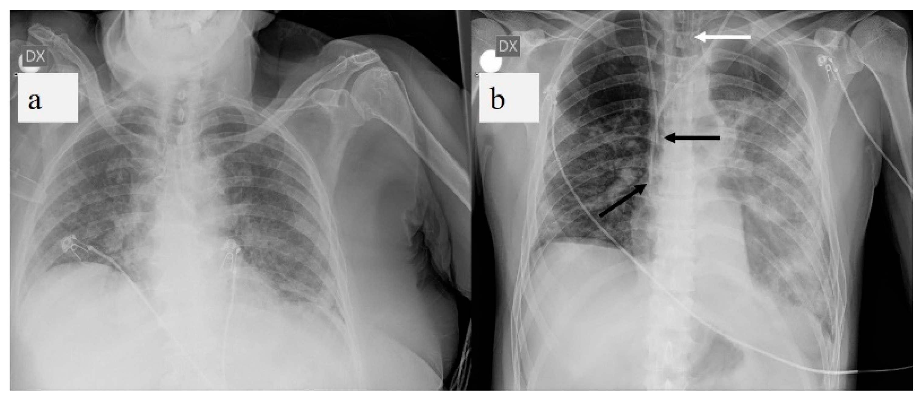
Diagnostics Free Full Text A Pictorial Review Of The Role Of Imaging In The Detection Management Histopathological Correlations And Complications Of Covid 19 Pneumonia Html
0 Response to "Characteristic Beam Patterns of X-ray Include Which of the Following"
Post a Comment